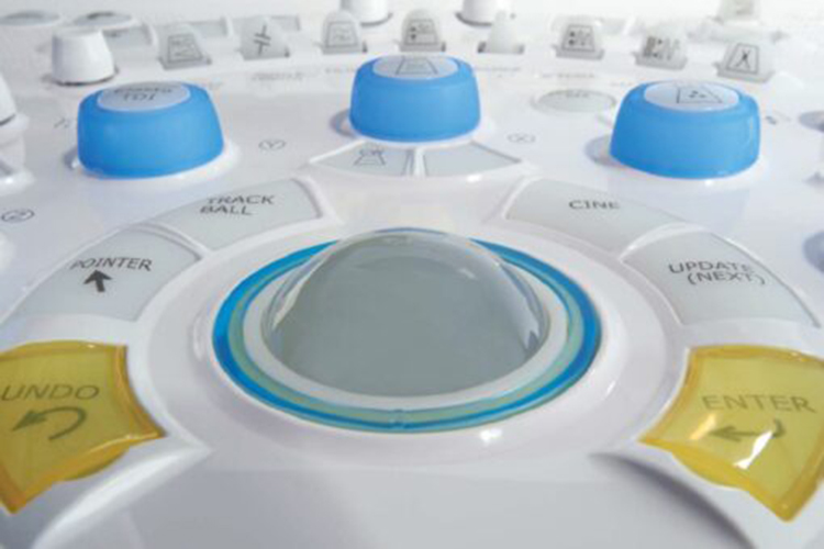
Hitachi HI VISION Preirus – Ultrasound System - USED
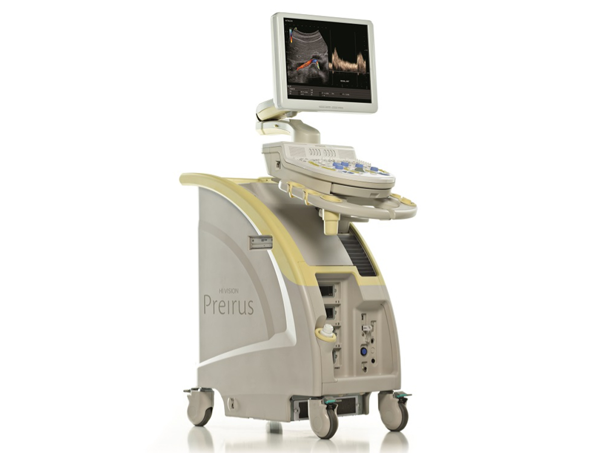
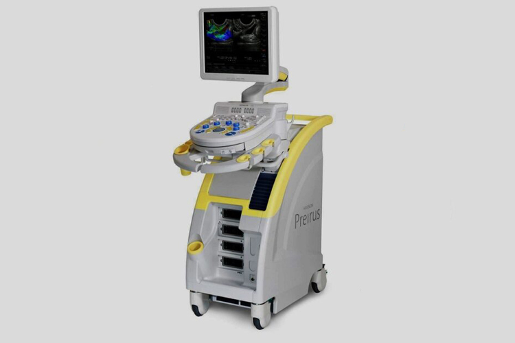
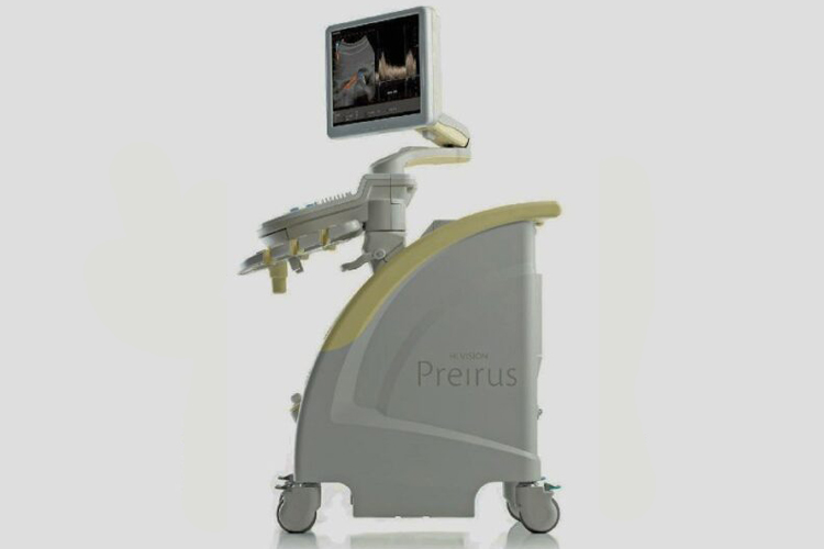
The Hitachi HI VISION Preirus is a high-performance ultrasound system designed for various medical imaging applications, including abdominal, cardiac, vascular, obstetric, gynecological, and musculoskeletal examinations. Known for its advanced imaging technologies, ergonomic design, and ease of use, this system is favored in clinical environments for producing high-resolution images with enhanced diagnostic accuracy.
Key Features:
Advanced Imaging Technologies:
- Hitachi's HI VISION technology: Provides high-definition image clarity with real-time tissue harmonics and advanced Doppler capabilities.
- Real-time Tissue Elastography (RTE): Assesses tissue stiffness, useful for detecting conditions like tumors.
- Smart 3D and 4D Imaging: Especially useful in obstetrics and gynecology for clear visualization of fetal anatomy.
Ergonomics and Usability:
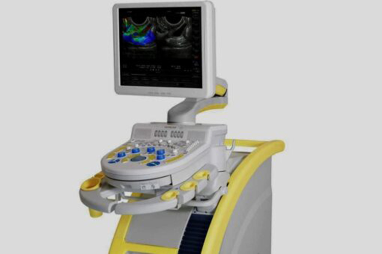
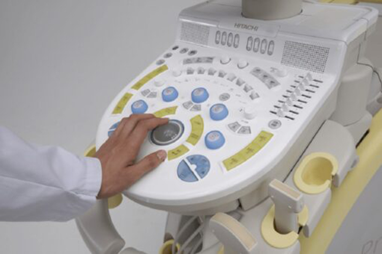
- Intuitive interface: Touchscreen controls with customizable options for different scanning modes.
- Compact and adjustable: The design includes a compact footprint and adjustable console for ease of use and operator comfort.
Transducer Compatibility: The system supports a wide variety of transducers, enabling a range of diagnostic applications, from routine scans to more specialized uses.
Doppler Imaging: The system offers color Doppler, power Doppler, and spectral Doppler capabilities for vascular studies and cardiac assessments.
Enhanced Workflow:
- Integration of DICOM for seamless connectivity with PACS systems.
- On-board reporting and archiving features.
Applications:
- Cardiology: Provides high-quality cardiac imaging with Doppler functionalities for assessing blood flow and heart conditions.
- Obstetrics & Gynecology: 3D/4D imaging allows detailed views of fetal development.
- Musculoskeletal: Offers detailed visualization of soft tissues, tendons, and joints.
- Vascular: Doppler technologies support comprehensive vascular assessments.
This system has been widely recognized for its imaging precision, making it a valuable tool for healthcare providers.
